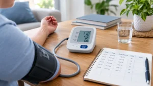


Sharp back pain by itself is usually muscular. Low urine output by itself is usually dehydration. Together, they describe a blocked kidney until proven otherwise, and blockage destroys kidney tissue on a clock. Pressure inside an obstructed kidney begins reducing filtration within hours. Function that returns almost completely after 24 hours of obstruction may not return after seven days.
That is why this specific combination belongs in an emergency department rather than a next-day clinic slot. A renal ultrasound and a bladder scan settle the question in under 30 minutes, and relieving pressure with a catheter, stent, or nephrostomy tube often restores urine flow the same visit. Southern Oklahoma Kidney Center manages the medical side of these cases through our nephrology practice in Ardmore.
Go Now If Any of These Apply
- Less than 400 mL of urine in 24 hours (under two standard cups), or nothing at all for 12 hours
- Flank pain plus fever above 101°F, chills, or shaking — a possible infected obstructed kidney
- Pain that comes in waves from the flank into the groin with vomiting
- A bladder that feels full while nothing comes out
- Visible blood or clots in the urine with reduced flow
- Confusion, low blood pressure, or a heart rate above 110 with any of the above
- One functioning kidney, a transplant, or existing CKD with new flank pain
An infected, obstructed kidney is the highest-mortality presentation in this group. Bacteria trapped behind a blockage seed the bloodstream, and antibiotics alone cannot fix it. Drainage within hours is the treatment.
Kidney Pain Versus Back Pain: How to Tell
| Feature | Kidney Origin | Musculoskeletal Origin |
|---|---|---|
| Location | Flank, below the ribs, beside the spine; often one side | Midline lower back, buttocks, or across the belt line |
| Pattern | Deep, constant, or wave-like colic; unrelieved by position | Worse with bending or lifting; eases with rest or heat |
| Radiation | Toward the groin, testicle, or labia | Down the back of the leg (sciatic pattern) |
| Associated signs | Fever, nausea, vomiting, blood in urine, reduced output | Muscle spasm, tenderness on touch, recent exertion |
| Effect of movement | Patient cannot find a comfortable position | Specific positions bring relief |
Renal colic classically makes patients pace; back strain makes them lie still. That single observation separates most cases at the bedside.
Why Urine Output Drops: Four Mechanisms
| Mechanism | Typical Story | Key Test | Fix |
|---|---|---|---|
| Bladder outlet obstruction | Enlarged prostate, medication effect, full-feeling bladder, dribbling | Bladder scan; residual over 300 mL | Urinary catheter, often immediate relief |
| Ureteral stone | Sudden colicky flank pain radiating to groin, blood in urine | Non-contrast CT or ultrasound | Stent or ureteroscopy; stones over 6 mm rarely pass alone |
| Bilateral obstruction or single-kidney blockage | Cancer, retroperitoneal disease, clots, prior surgery | Renal ultrasound showing hydronephrosis | Nephrostomy tube drainage |
| Severe volume depletion or shock | Heat exposure, vomiting, diarrhea, bleeding, sepsis | Metabolic panel, blood pressure, FENa | IV fluids and treatment of the source |
Volume depletion produces low output without the full-bladder sensation. Obstruction produces low output with pressure and pain. Both raise creatinine, so labs alone cannot separate them — imaging does. Stone-prone patients should review hydration and kidney stone risk.
The Obstruction Clock
Kidney damage from blockage follows a predictable timeline. This framework does not exist on most competing pages, and it explains why “waiting until Monday” carries a real cost.
| Duration of Complete Obstruction | What Happens Inside the Kidney | Expected Recovery After Drainage |
|---|---|---|
| Under 24 hours | Pressure rises; filtration falls; tissue intact | Near-complete return of function |
| 1–7 days | Tubular injury begins; urine concentrating ability drops | Substantial recovery, often over days to weeks |
| 1–4 weeks | Progressive tubular atrophy and early fibrosis | Partial recovery; residual CKD common |
| Beyond 6–12 weeks | Cortical thinning and permanent scarring | Limited recovery; the kidney may not return to function |
Add infection to any row and the timeline compresses to hours. Add a single functioning kidney and every row becomes an emergency.
Partial obstruction behaves differently. Urine still passes, output may look normal, and pain can be mild or intermittent — yet pressure quietly damages tissue for weeks. Patients with a known stone, prior stent, or prostate enlargement who notice a weakening stream, rising creatinine on routine labs, or one-sided ache lasting more than a few days need an ultrasound even without dramatic symptoms. Silent partial obstruction accounts for a meaningful share of unexplained kidney function loss found on annual bloodwork.
Sepsis: The Complication That Moves Fastest
An obstructed kidney that becomes infected produces urosepsis, and urosepsis kills faster than kidney failure. Recognize it by fever or a temperature below 96.8°F, heart rate above 90, breathing rate above 20, confusion, and systolic blood pressure under 100 mmHg. Any two of those with flank pain justify an ambulance rather than a car.
Emergency management runs on a clock: blood cultures drawn, broad-spectrum antibiotics started within the first hour, IV fluids for perfusion, and urgent drainage of the blocked system. Antibiotics without drainage fail because the drug cannot reach bacteria behind a closed obstruction. Ask directly whether drainage is planned and when. Diabetic patients, transplant recipients, and anyone on immunosuppression progress from fever to septic shock in a compressed timeframe, which is why the same symptoms warrant faster action in those groups.
What Happens at Mercy Hospital Ardmore
The First 60 Minutes
Expect a focused sequence: vital signs, a basic metabolic panel with creatinine and potassium, urinalysis, a bladder scan, and a renal ultrasound. Fever prompts blood cultures and a lactate level. A non-contrast CT of the abdomen and pelvis identifies stones with high accuracy and avoids contrast dye that would further stress an injured kidney.
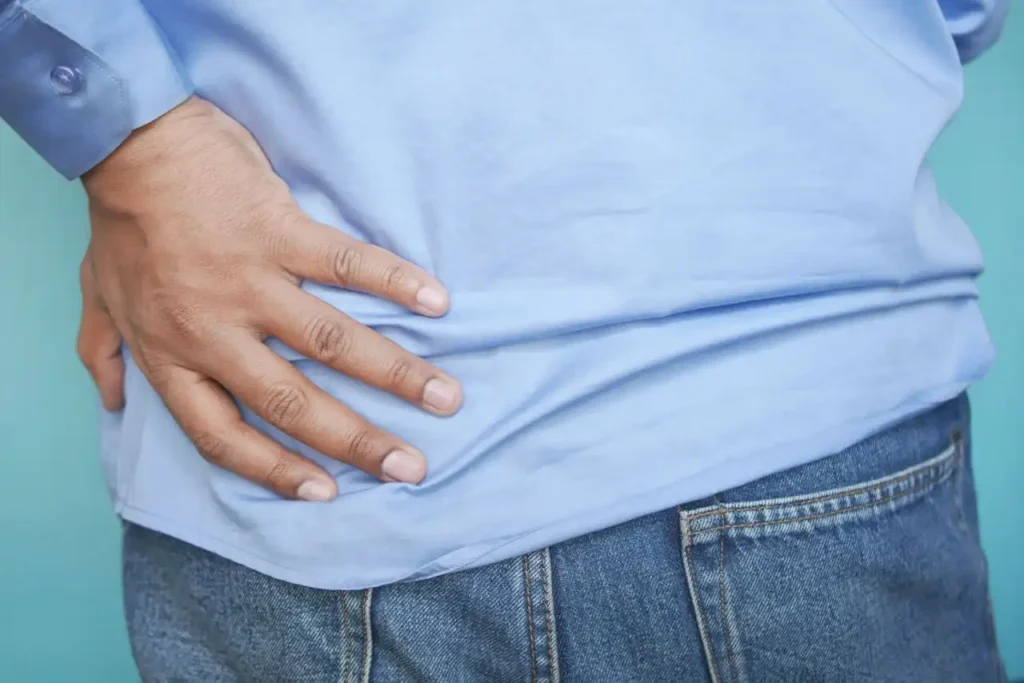
Decompression Options
- Urethral catheter: resolves bladder outlet obstruction at the bedside; output of over a liter immediately after placement confirms the diagnosis
- Ureteral stent: placed by a urologist to bypass a stone or stricture
- Percutaneous nephrostomy: a drain placed through the skin into the kidney when a stent cannot pass or infection demands rapid drainage
Watch for post-obstructive diuresis afterward. Once pressure releases, some patients produce several liters of urine over the next 24 hours and lose sodium, potassium, and magnesium rapidly. This phase needs monitored fluid and electrolyte replacement, not discharge. Kidney and urinary specialists share this work — see how urologists and nephrologists coordinate care.
Pain Control Without Kidney Harm
Renal colic pain is severe and treatable. Emergency clinicians typically use IV ketorolac, an NSAID, because it works well for stone pain — but ketorolac is nephrotoxic and inappropriate when creatinine is already rising or when obstruction is suspected in a single kidney. Say your creatinine number out loud and ask whether an opioid or IV acetaminophen is safer for your situation. Requesting the safer option costs nothing and prevents a second injury layered on the first.
Southern Oklahoma Risk Context
| Regional Factor | Why It Raises Obstruction and AKI Risk Here | Countermeasure |
|---|---|---|
| Summer heat and outdoor labor | Concentrated urine promotes stone formation; Oklahoma sits near the southern stone belt | Target pale-yellow urine; increase intake before and during shifts |
| High red meat and sodium intake | Raises urinary calcium and uric acid saturation | Lower sodium; add citrus for urinary citrate |
| Men over 60 in rural areas | Untreated prostate enlargement progresses to retention | Report weak stream and nighttime urination before retention occurs |
| Anticholinergic and decongestant use | Cold medicines and bladder drugs precipitate acute retention | Review medications after any retention episode |
| Distance from specialty urology | Delays stent placement after hours | Present early; transfer decisions are faster before sepsis develops |
After the Emergency: Preventing the Repeat
Discharge is the midpoint, not the end. Book a follow-up creatinine within one to two weeks and again at three months, because obstruction that lasted more than a day leaves measurable scarring in a meaningful share of patients. Stone formers need a 24-hour urine collection to identify the metabolic driver — calcium oxalate, uric acid, cystine, or struvite — since each has a different prevention plan. Patients with recurrent stones should read when to see a nephrologist for kidney stones, and everyone leaving the hospital should understand their numbers through our guide to kidney function tests.
Ongoing stone management, metabolic testing, and medication adjustment run through our kidney stone program.
Frequently Asked Questions
How long can a kidney stay blocked before permanent damage?
Meaningful injury begins within 24 hours and worsens over the following week. Complete obstruction beyond several weeks often leaves permanent function loss. Infection shortens the window to hours.
Can back pain alone mean kidney failure?
Rarely. Kidney failure usually presents with fatigue, swelling, nausea, and reduced urine rather than pain. Pain plus reduced output is the combination that matters.
How much urine is too little?
Under 400 mL in 24 hours is oliguria. Under 100 mL is anuria and requires immediate emergency evaluation. For a 175-pound adult, hourly output should stay near 40 mL.
Will drinking more water flush out a blockage?
No. Extra fluid behind a complete blockage raises pressure and worsens pain and injury. Drink normally and seek imaging.
Is a kidney stone always the cause?
No. Prostate enlargement, blood clots, tumors, scar tissue, and pelvic surgery complications all obstruct. Imaging identifies the level of the blockage, which determines the drainage method.
What if the pain stops but I am still not urinating?
Treat that as more dangerous, not less. Pain often fades once the kidney stops producing urine behind the blockage. Loss of pain with persistent low output suggests the kidney has shut down and needs urgent imaging.
Can both kidneys be blocked at once?
Yes. Bladder outlet obstruction, pelvic tumors, and retroperitoneal disease block both drainage systems simultaneously and cause rapid creatinine rise with almost no urine output. This scenario produces the sharpest lab changes and needs same-day drainage.
Talk to a Nephrologist in Ardmore
Southern Oklahoma Kidney Center evaluates post-obstruction kidney function, stone metabolism, and acute kidney injury from 2611 Crossroads Drive, Ardmore, OK 73401, with clinics in Ada and Gainesville. Call (580) 223-8614 or book a follow-up visit after any emergency department visit for flank pain with low urine output. For active symptoms with fever or no urine output, go to the emergency department first, then contact our office for follow-up.
Medically reviewed by the nephrology team at Southern Oklahoma Kidney Center. Educational content only. Call 911 for emergencies.