
You passed a kidney stone. The pain is gone, the ER bill has arrived, and someone told you to “drink more water.” That advice is fine, but it is incomplete.
About half of people who pass one stone will form another within 5 to 10 years. The best way to break that cycle is to find out exactly what your stone is made of, then match your diet and medicine to that chemistry. Advice that prevents one stone type can make another type worse.
Why Stone Composition Matters
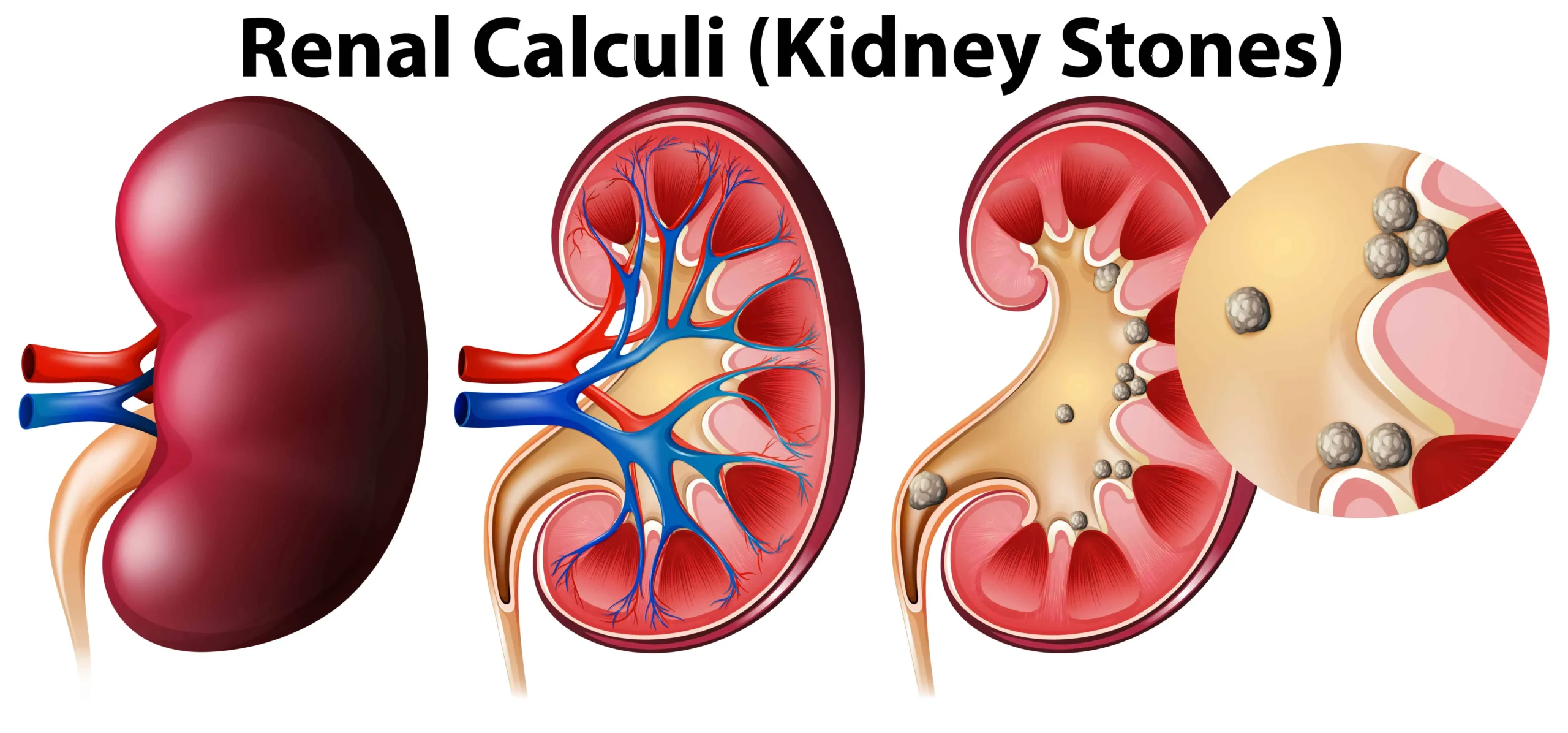
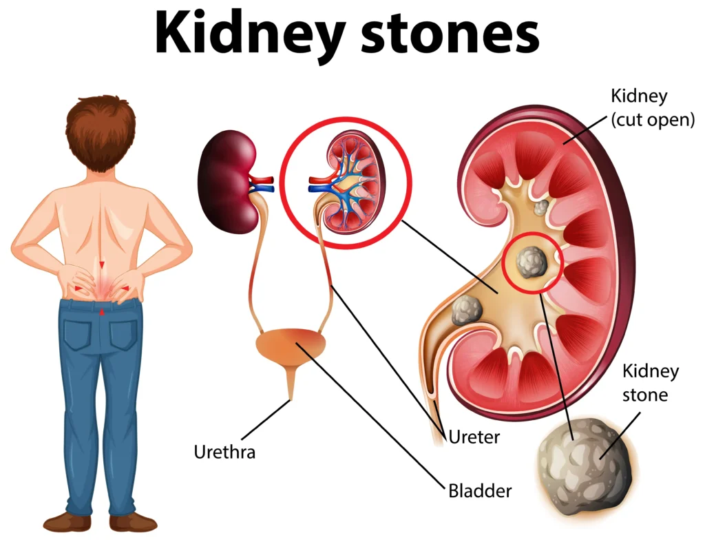
A kidney stone is a crystal that grows when urine holds more of a mineral than it can keep dissolved. Doctors call this supersaturation. Different minerals crystallize under different conditions:
- Some form in acidic urine, others in alkaline urine.
- Some come from diet, others from genetics, infection or medicines.
- Some show up clearly on X-ray, others are almost invisible.
Treating every stone the same is like treating every infection with the same antibiotic. Our kidney stone (nephrolithiasis) management program starts with identifying the type.
The Main Types of Kidney Stones
| Stone Type | Share of Stones | Urine Conditions | Common Causes | Shows on Plain X-ray? |
| Calcium oxalate | About 70% to 80% | Any pH | Low urine volume, high urine calcium or oxalate, low citrate | Yes |
| Calcium phosphate | About 10% to 15% | Alkaline (pH above 6.5) | Renal tubular acidosis, overactive parathyroid, some medicines | Yes |
| Uric acid | About 5% to 10% | Acidic (pH below 5.5) | Obesity, type 2 diabetes, gout, high animal protein diet | No |
| Struvite | About 1% to 5% | Alkaline | Urinary infection with urease-producing bacteria | Yes |
| Cystine | Under 2% | Acidic or neutral | Inherited cystinuria | Faintly |
| Drug-induced | About 1% | Varies | Topiramate, some HIV medicines, triamterene, certain antibiotics | Varies |
Calcium Oxalate Stones
These are the most common by far. Two forms exist: monohydrate, which is very hard, and dihydrate, which breaks more easily during lithotripsy. Low urine volume is the biggest driver. High urine calcium, high oxalate and low citrate add to the risk. Citrate is a natural stone blocker, and many stone formers simply do not make enough of it.
Calcium Phosphate Stones
These form in alkaline urine. A pattern of repeated calcium phosphate stones should prompt a check for distal renal tubular acidosis or primary hyperparathyroidism. The seizure and migraine drug topiramate raises the risk too.
Uric Acid Stones
The key problem here is acidic urine, not always high uric acid. People with obesity, insulin resistance and type 2 diabetes tend to make very acidic urine. Here is the good part: uric acid stones are the only common type that can dissolve with medicine. Raising urine pH to 6.0 to 6.5 with potassium citrate can shrink them without surgery.
Struvite (Infection) Stones
Struvite is magnesium ammonium phosphate. It forms when bacteria such as Proteus split urea and turn urine alkaline. These stones can grow fast and fill the kidney in a branching “staghorn” shape. They are more common in women and in people with catheters or neurogenic bladder. Antibiotics alone will not cure them. The whole stone usually has to come out.
Cystine Stones
Cystinuria is a genetic condition that lets the amino acid cystine spill into the urine. Stones often begin in childhood or the teen years and come back often. Treatment needs very high fluid intake, urine alkalinization and sometimes a cystine-binding drug such as tiopronin.
How Doctors Find Out What Your Stone Is Made Of
Stone Analysis
If you pass a stone, catch it. Urinate through a strainer for a few days after an episode, let the stone dry and bring it to your appointment. Labs use infrared spectroscopy or X-ray diffraction to identify each mineral layer. Many stones are mixed, such as calcium oxalate with a calcium phosphate core, and that mix changes the plan.
Had a stone removed by ureteroscopy? Ask your urologist to send fragments for analysis. Our article on how urologists and nephrologists work together explains how that handoff works.
CT Scan Clues
A CT scan measures stone density in Hounsfield units. Uric acid stones usually read under 500, while calcium stones often read above 1,000. Dual-energy CT can separate uric acid from calcium stones before the stone ever leaves your body.
The 24-Hour Urine Test
Stone analysis tells you what formed. A 24-hour urine collection tells you why. You collect every drop of urine for one full day on your normal diet, ideally at least 6 weeks after a stone episode. The lab measures:
- Total urine volume (goal is at least 2.5 liters a day)
- Calcium, oxalate, citrate and uric acid
- Sodium, which pushes more calcium into urine
- pH, magnesium, phosphate and sulfate (a marker of animal protein intake)
- Creatinine, to confirm the collection was complete
The report calculates supersaturation for each stone type. That turns vague advice into specific targets.
Blood Tests
Calcium, parathyroid hormone, uric acid, bicarbonate and kidney function round out the workup. Repeated stones can quietly harm kidney function, so we track creatinine over time.
Prevention Plans by Stone Type
| Stone Type | Diet Changes | Medicines Often Used | Urine Target |
| Calcium oxalate | Normal calcium (1,000 to 1,200 mg) with meals, low sodium, limit high-oxalate foods, avoid high-dose vitamin C | Thiazide diuretic for high urine calcium, potassium citrate for low citrate | Volume 2.5 L or more, calcium and oxalate in range, citrate above normal |
| Calcium phosphate | Low sodium, moderate calcium | Thiazide; citrate used with care since it raises pH | Avoid pH above 6.5 |
| Uric acid | Less red meat, organ meat and shellfish; weight loss | Potassium citrate; allopurinol if uric acid is high | pH 6.0 to 6.5 |
| Struvite | Fluids | Complete stone removal, targeted antibiotics, sometimes acetohydroxamic acid | Sterile urine |
| Cystine | Low sodium, less animal protein | Potassium citrate, tiopronin | Volume 3 L or more, pH above 7.0 |
The Calcium Myth
Many patients cut out dairy after a calcium oxalate stone. That usually backfires. Calcium in food binds oxalate in the gut so less of it reaches the urine. Cutting calcium raises oxalate absorption and weakens bones. Keep dietary calcium normal and pair it with meals.
A renal dietitian can build a realistic menu around your results. Learn more about the role of a renal dietitian in kidney care.
Stone Risk Factors in Southern Oklahoma and North Texas
This region sits on the western edge of what doctors call the “stone belt,” where hot summers and southern eating habits push stone rates higher.
| Local Factor | Why It Raises Stone Risk | What to Do |
| Summer heat and long outdoor shifts | Sweat loss concentrates urine for hours | Carry a marked water bottle; aim for pale yellow urine all day |
| Brewed iced tea and sweet tea | Black tea is high in oxalate; sugar raises urine calcium | Cut back, add lemon, alternate with water |
| Salty processed and fried foods | Sodium pulls calcium into the urine | Keep sodium under 2,300 mg a day, lower if advised |
| High rates of obesity and diabetes | Acidic urine favors uric acid stones | Weight control, check urine pH |
| Limited restroom access at work | People drink less to avoid breaks | Plan fluid timing around breaks |
Work routines matter more than most people think. See our article on habits at work that may affect kidney health. For hydration specifics, read hydration and kidney stones.
Cost of Prevention vs. Cost of Another Stone
| Item | Prevention Path | Repeat Stone Path |
| Testing | Stone analysis and 24-hour urine, often repeated yearly | ER visit with CT scan |
| Treatment | Low-cost generic medicines such as thiazides or potassium citrate | Possible ureteroscopy, stent or lithotripsy |
| Time lost | One clinic visit and one collection day | Days to weeks of pain and missed work |
| Kidney impact | Protects long-term function | Repeated blockages and procedures can scar the kidney |
For most recurrent stone formers, a single metabolic workup costs less than one emergency visit.
When to Seek Urgent Care
Go to the ER if you have stone pain with fever or chills, vomiting that stops you from keeping fluids down, or very little urine. A blocked, infected kidney is an emergency. Our article on when sharp back pain and barely any urine mean an ER visit explains the red flags.
Frequently Asked Questions
Can I tell my stone type by how it looks?
Not reliably. Color and shape give hints, but only lab analysis confirms the mineral makeup.
Should I avoid all high-oxalate foods?
Only if your 24-hour urine shows high oxalate. Spinach, rhubarb, almonds and beets are the biggest sources. Most people just need moderation, not elimination. Our list of kidney-friendly foods can help with swaps.
Does lemon water help?
Lemon juice adds a small amount of citrate. It helps some calcium stone formers but does not replace prescribed potassium citrate when citrate is very low.
Who should I see, a urologist or a nephrologist?
A urologist removes stones. A nephrologist finds out why they form and works to stop the next one. Read when to see a nephrologist for kidney stones for more detail.
Stop the Next Stone Before It Starts
If you have had two or more stones, a single large stone, a stone before age 25, or one kidney, you should get a full metabolic workup. Bring your stone, any CT reports and a list of your medicines.
Southern Oklahoma Kidney Center sees stone patients in Ardmore and Ada, OK, and at our Gainesville, Texas kidney clinic. Call (580) 223-8614 or request an appointment to start your stone prevention plan.
This article is for education only and does not replace advice from your own doctor.